Instrumentation
| Instrument | Contact | Description |
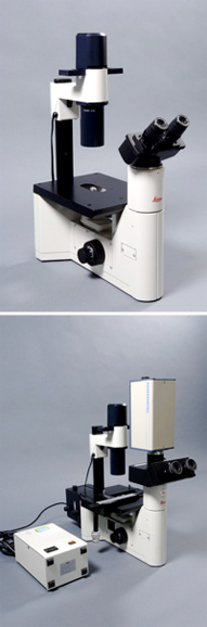
Inverse Leica DMIL |
UZH, MUL |
Brightfield and phasecontrast; Objectives: 4x, 10x, 20x, 40x; the master microscope is equiped with epifluorescence and a c-mount adapter. Fluorescence: Blue exitation (FITC, Cy2, Alexa 488); green exitation (TRITC, Cy3, Alexa 546). Kostenlos für Kurse der Universität oder Kurse, an welchen die Universität beteiligt ist. |
Zeiss Axiolab |
UZH, MUL |
Brightfield and phasecontrast; Epifluorescence: UV-exitation (Dapi, Hoechst); blue exitation (FITC, Cy2, Alexa 488); green exitation (TRITC, Cy3, Alexa 546). Objectives: 10x, 40x, 100x, Oelimmersion. The master microscope is equiped with better objectives and a c-mount adapter. Kostenlos für Kurse der Universität oder Kurse, an welchen die Universität beteiligt ist. |
FACScanto |
UZH, Physiology or |
Flow cytometer with two lasers (488 and 633 nm) and autosampler (carousel) and allows you to measure up to six colors. Registration is only possible if you are an experienced FACS user or if you have taken a course by BD Bioscience. |
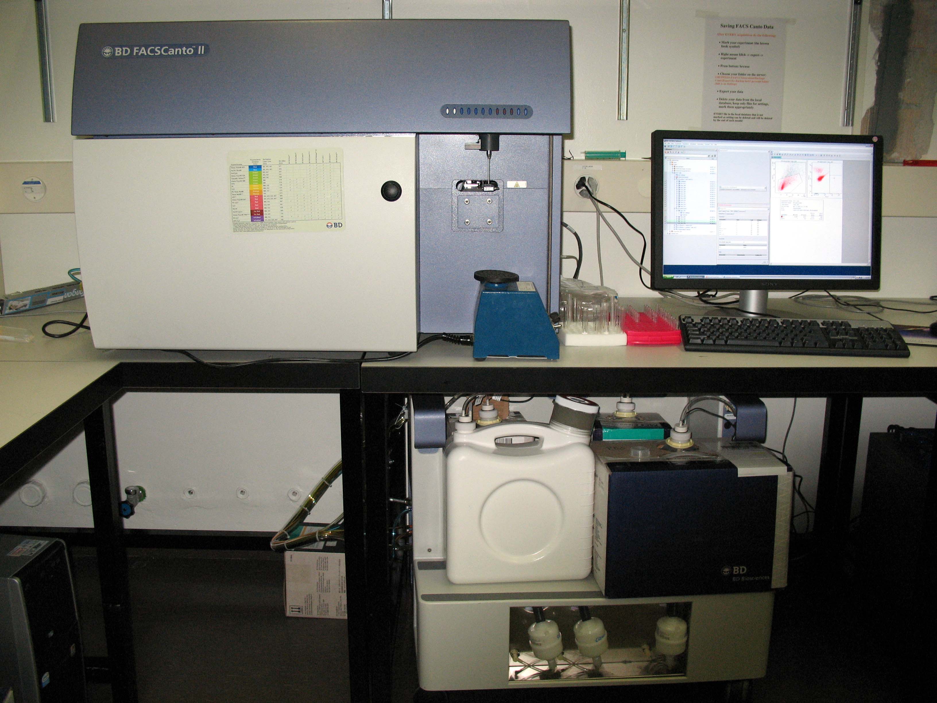
FACScanto II |
UZH, exp. Immunology |
The FACScanto II has three lasers (405, 488 and 633 nm) and allows you to measure up to eight colors. Registration is only possible if you are an experienced FACS user or if you have taken a course by BD Bioscience. |
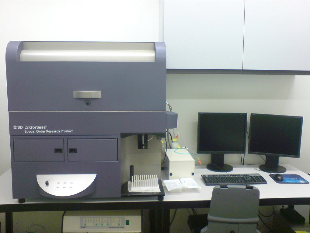
LSRII Fortessa |
UZH, exp. Immunology |
High-end BD SORP flow cytometer with 4 lasers and 18 measureable parameters (usually 2 light-scatter signals and 16 fluorescent signals). Violet laser (405 nm) is coupled to 6 PMTs, blue laser (488 nm) is coupled to 3 PMTs, yellow-green laser (561 nm) is coupled to 5 PMTs and red laser (640 nm) is coupled to 3 PMTs. |
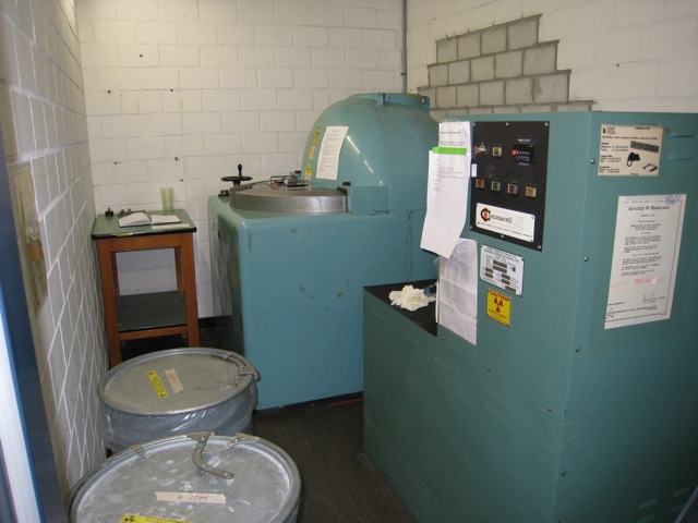
Irradiation, Cs or Co source |
USZ, Immunology Lukas Heeb lukas.heeb@usz.ch 076 517 91 11 |
The Co source is mainly used for irradiation of cells. The max size of your irradiation goods is limited by the size of the chamber which has the following measures: H 13.5cm, W 16 cm, B 11 cm. You can irradiate anything that fits into this chambers and is compliant with the facility regulations and the SOP. The Cs source is meant to irradiate small animals (mice). |
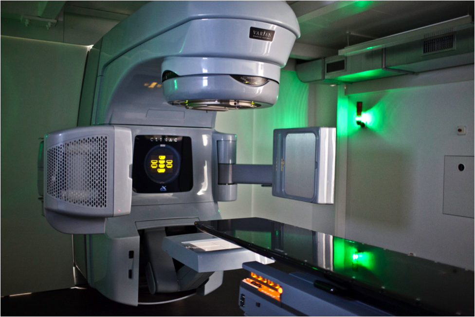
Irradiation, Linear accelerator |
UZH, Vetsuisse Faculty Division of Radiation Oncology DACVR (Radiation Oncology), DipECVDI (add Rad Oncol) |
The linear accelerator is mainly used for irradiation of pet animals (cats and dogs) in clinical and research setting. However, researchers are welcome and encouraged to use the machine also for irradiation of cells or laboratory animals. Due to the high technical complexity of the machine, treatment will always be performed by our staff. Radiation dose will be calculated at least one day prior to the session by a radiation oncologist and approved by a medical physicist. Field sizes from 2 cm to at least 25 cm can be treated. The radiation dose will be applied in electron or photon mode, depending on the object irradiated (e.g. mouse, cell culture, etc.). The time needed for the application of a fraction is fast but dose dependent. Available energies and treatment modes: Photons 6 MeV |
IVIS 200 Bioluminescence |
USZ, Immunology Laura Bürgi laura.buergi@usz.ch 044 634 15 03 |
The Xenogen IVIS Imaging System 200 is a high-sensitivity, low noise, in vivo imaging technology platform that allows non-invasive visualization and tracking of cellular and genetic activity within a living organism, in real time. Registration is only possible if you are an experienced IVIS user and if you have been given specific instructions by the IVIS laboratory personnel for this specific machine. |
Small-animal image-guided radiotherapy platform |
UZH Irchel, Physiologie – Applied Radiobiology |
The small-animal image-guided radiotherapy platform PXI 225 CX is a machine for the precise irradiation of mice and rats. An integrated cone beam-CT acquires high-resolution CT scans of the tissues and bones. These scans are subsequently loaded to a treatment-planning software, where the researcher can construct an individual treatment plan for the animal. The organs or structures of interest can be irradiated from different angles with X-ray beam diameters between 3mm and 40mm. The beam energy is at 225kV and the dose rate is 3 Gy/min. In addition, the platform also contains an optical camera for the acquisition of bioluminescence. Note: The platform can only be used under supervision of a trained employee from the group of applied radiobiology. |
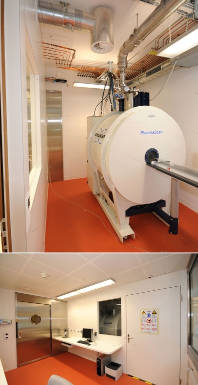
Small animal MRI |
USZ, BZL Udo Ungethüm 044 255 20 40 oder Conny Waschkies 044 253 0470 |
Our preclinical MRI Facility is equipped with a Bruker 4.7T PharmaScan® MRI system designed for high throughput preclinical imaging and biomedical and molecular imaging applications of small animals and tissue samples up to 6 cm in diameter. It features routine MRI sequences optimized for small rodents such as rats and mice and is operated by ParaVision® 5 and 6 software packages for data acquisition, reconstruction, analysis and visualization. The system consists of an actively shielded magnet and high-performance BGA-9 gradient system (300mT/m gradient strength and 3300 T/m/s slew rate), optimized for small animal imaging) interfaced to an AVANCETM III HD MRI RF spectrometer enabling MRI and MRS applications. Our RF coil landscape consists of two linearly polarized volume resonators for whole body and organ specific volume imaging in mice and rat (ID 38mm and ID 60mm), a quadrature transmit volume resonator (ID 72mm) and quadrature surface coil designed for imaging of mouse brain. |
UZH, Vet-Pathologie |
Automated Arcturus XT Laser Microdissection Instrument based on an inverted Nikon Eclipse Ti-E motorized Microscope. The system is equipped with an UV-cutting laser and an Infrared Laser for capturing dissected material. Objectives: 2x, 10x, 40x, 63x dry, 63x oil for epifluorescence, phase contrast and differential interference contrast. | |
| Bruker 7T PharmaScan® MRI system | High throughput preclinical imaging of small animals and tissue samples up to 6 cm in diameter; operated by ParaVision®360 software packages for data acquisition, reconstruction, analysis and visualization; high-field actively shielded magnet; high-performance BGA-9S HP gradient system (760mT/m gradient strength, 6800T/m/s maximum slew rate); RF coil landscape supports wide range of applications (neuro, cardiac, abdominal), 1H/23Na volume coil for sodium imaging; MRI sequences optimized for small rodents (routine anatomical, fMRI, DCE, ASL workflow, perfusion, diffusion, angiography, water-fat imaging, UTE, ZTE, UTE3D, 1H MR spectroscopy); added value by dynamic shimming capability, B1 optimization, chemical shift correction; IntraGate (Self-gating) cardiac imaging and IntraGateUTE for flow-artefact free cardiac MRI Equipment & Infrastructure: isoflurane anesthesia system, warm water bath for temperature control, physiological monitoring (body temperature, respiration, ECG) and gating system (SA Instruments Inc., NY) |